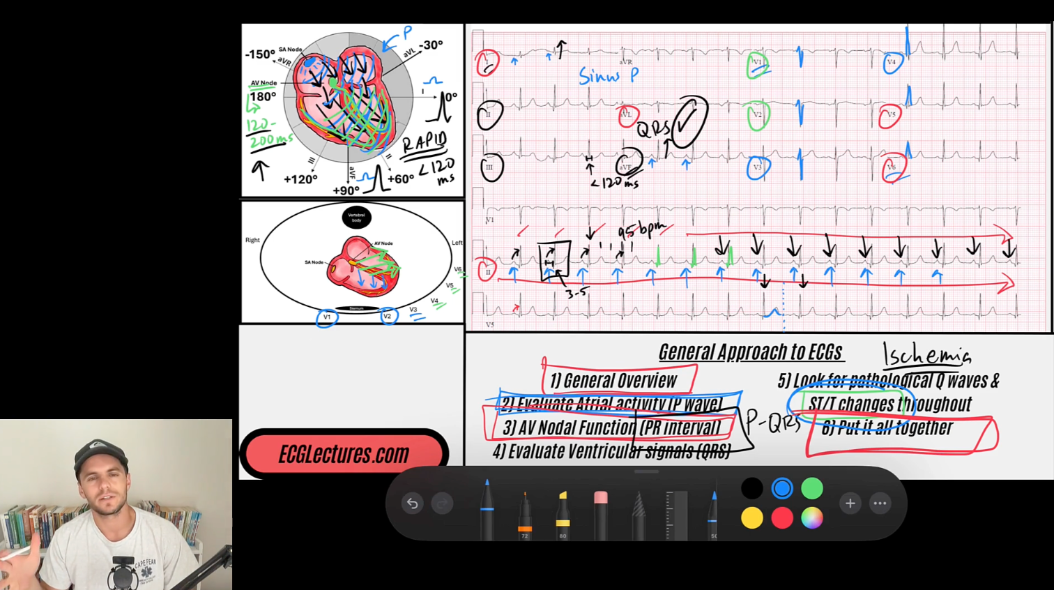
THE ECG BLOG WITH REID
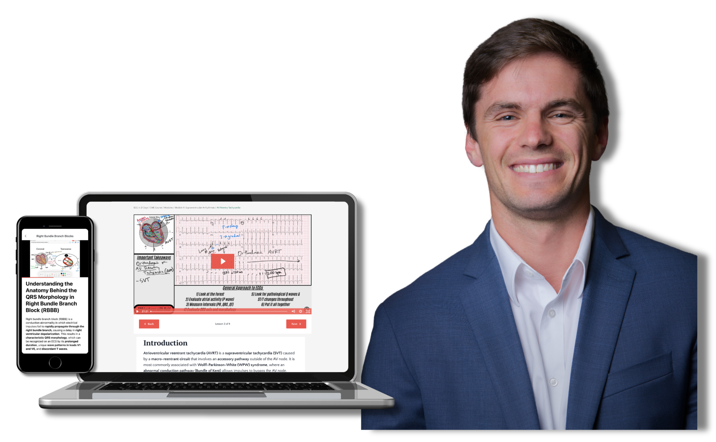
Open Access ECG Lectures & Blog Posts By Reid
Click below to learn more.
Rate-Related Wenckebach Phenomenon
Dec 09, 2025
Group Beating on ECG: Differentiating the Mechanisms
Dec 08, 2025
Mechanism of Delta Waves: Depolarization Fusion
Dec 07, 2025
Isorhythmic AV Dissociation from Sinus Arrhythmia
Dec 06, 2025
Atrial-Paced, Ventricular-Sensed Rhythms
Dec 04, 2025
Atrial-Sensed, Ventricular-Paced Rhythms
Dec 03, 2025
Two Diagnoses, One Tracing: A Case Review
Dec 02, 2025
The Anatomy Behind the P-Wave Terminal Force in V1
Dec 01, 2025
P-Wave Morphology in Premature Atrial Contractions
Nov 30, 2025
The Pseudo-r’ Wave in AVNRT
Nov 28, 2025