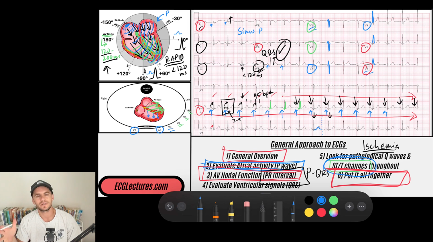
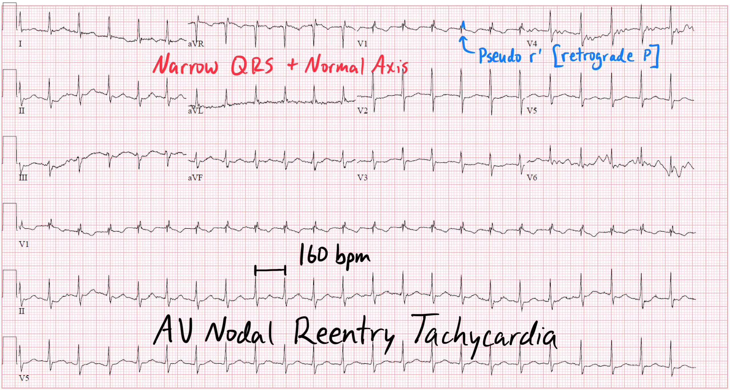
The Pseudo-r’ Wave in AVNRT
Nov 28, 2025One of the hallmark ECG clues for typical AV nodal reentrant tachycardia (AVNRT) is the appearance of a small, sharp pseudo-r’ deflection in lead V1. Although it looks like a tiny terminal R’ wave, it is not part of ventricular depolarization. Instead, it represents retrograde atrial activation occurring immediately after ventricular depolarization.
Unfamiliar with AVNRT? Read more & watch the AVNRT blog post here.
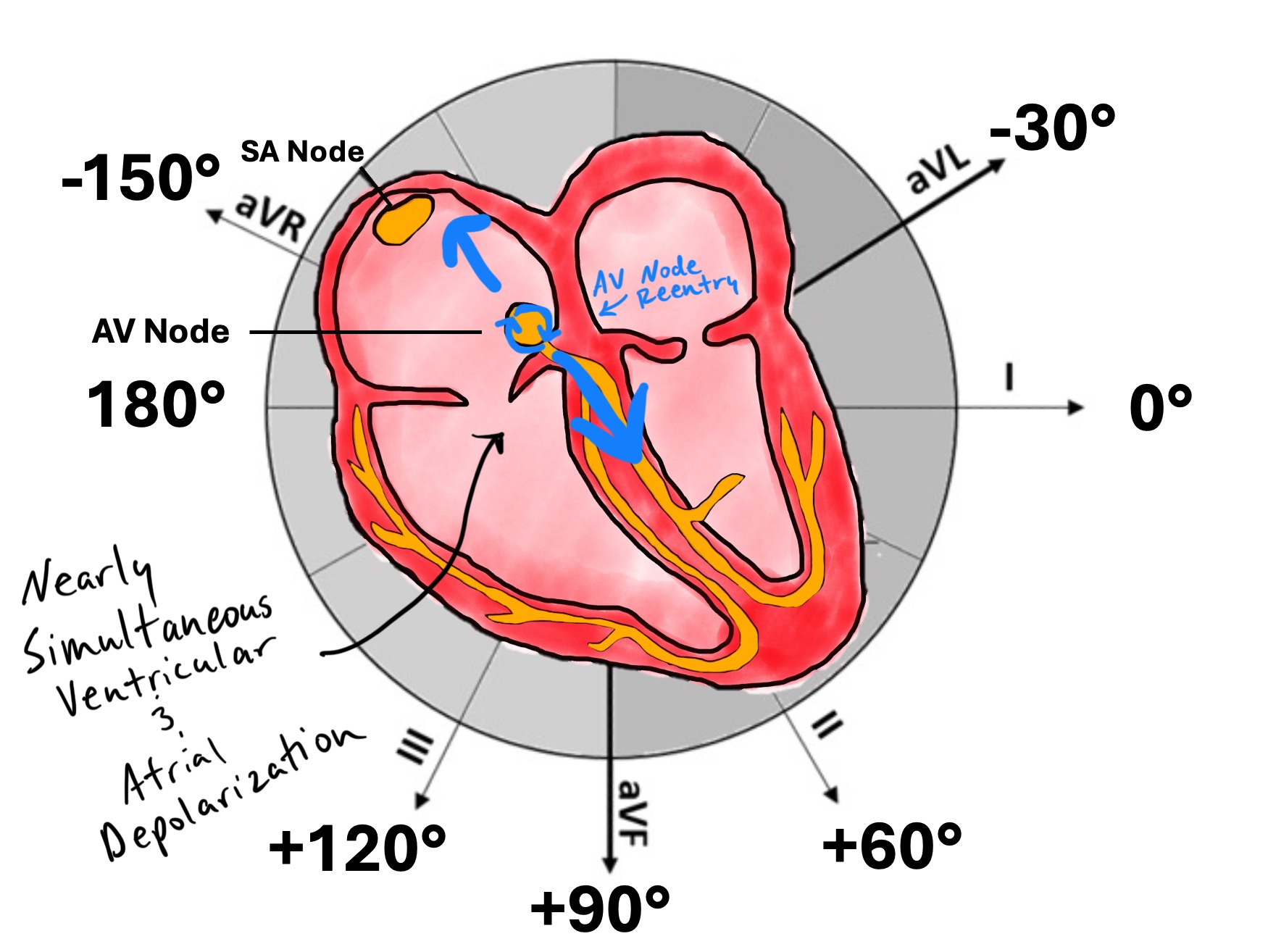
1. The Anatomical Basis: Dual Pathways of the AV Node
In typical AVNRT, the reentrant loop forms within the AV node and perinodal tissue, utilizing two distinct conduction pathways:
-
Slow pathway: Posterior–inferior, located near the coronary sinus ostium
-
Fast pathway: Anterior–superior, located near the compact node
During sinus rhythm:
-
Atrial impulses preferentially travel down the fast pathway.
During AVNRT:
-
The impulse travels anterograde through the slow pathway (slow conduction)
-
Then retrograde through the fast pathway (rapid conduction back to the atria)
This produces a near-simultaneous activation of the ventricles and atria.
2. Why the Retrograde P Wave Gets Hidden
Because retrograde conduction occurs rapidly through the fast pathway, the atria depolarize:
**• Very shortly after the ventricles depolarize
• With the atria activated from the septum upward**
This timing means that the retrograde P wave falls inside or immediately after the QRS complex, where it becomes visually obscured.
But depending on the lead orientation, part of that hidden P wave becomes visible — and that’s where the pseudo-r’ comes from.
Enjoy ECG Lectures with Reid? Here is a special gift from Reid
100 High Yield Annotated ECGs
Click below to download this free resource.