The Septal R Wave in V1 and V2: Anatomy, Physiology, and Clinical Relevance
Oct 30, 2025Join ECG With Reid Academy & master ECG interpretation with my accredited curriculum ⤵️
-->https://www.skool.com/ecgwithreid
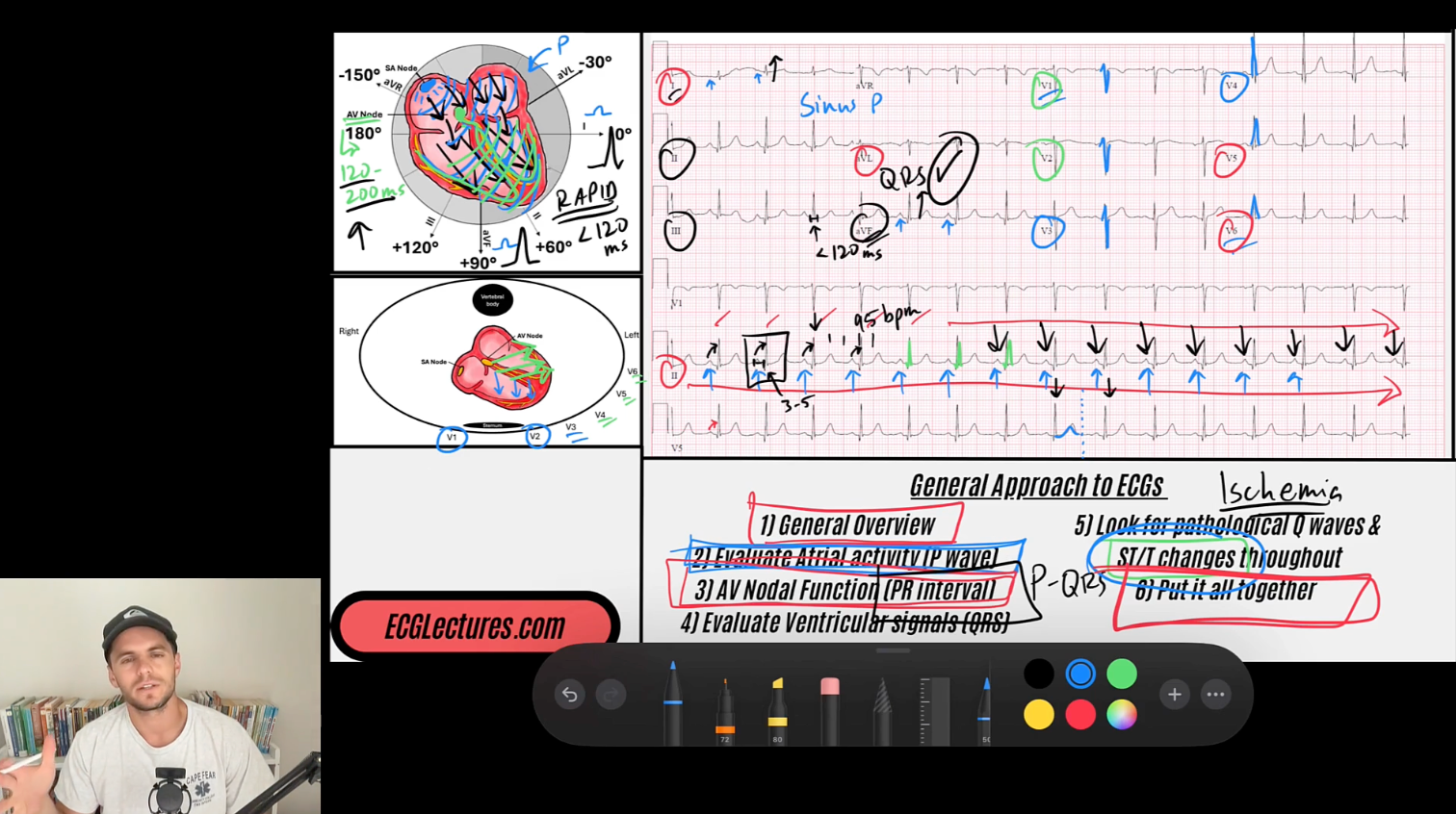
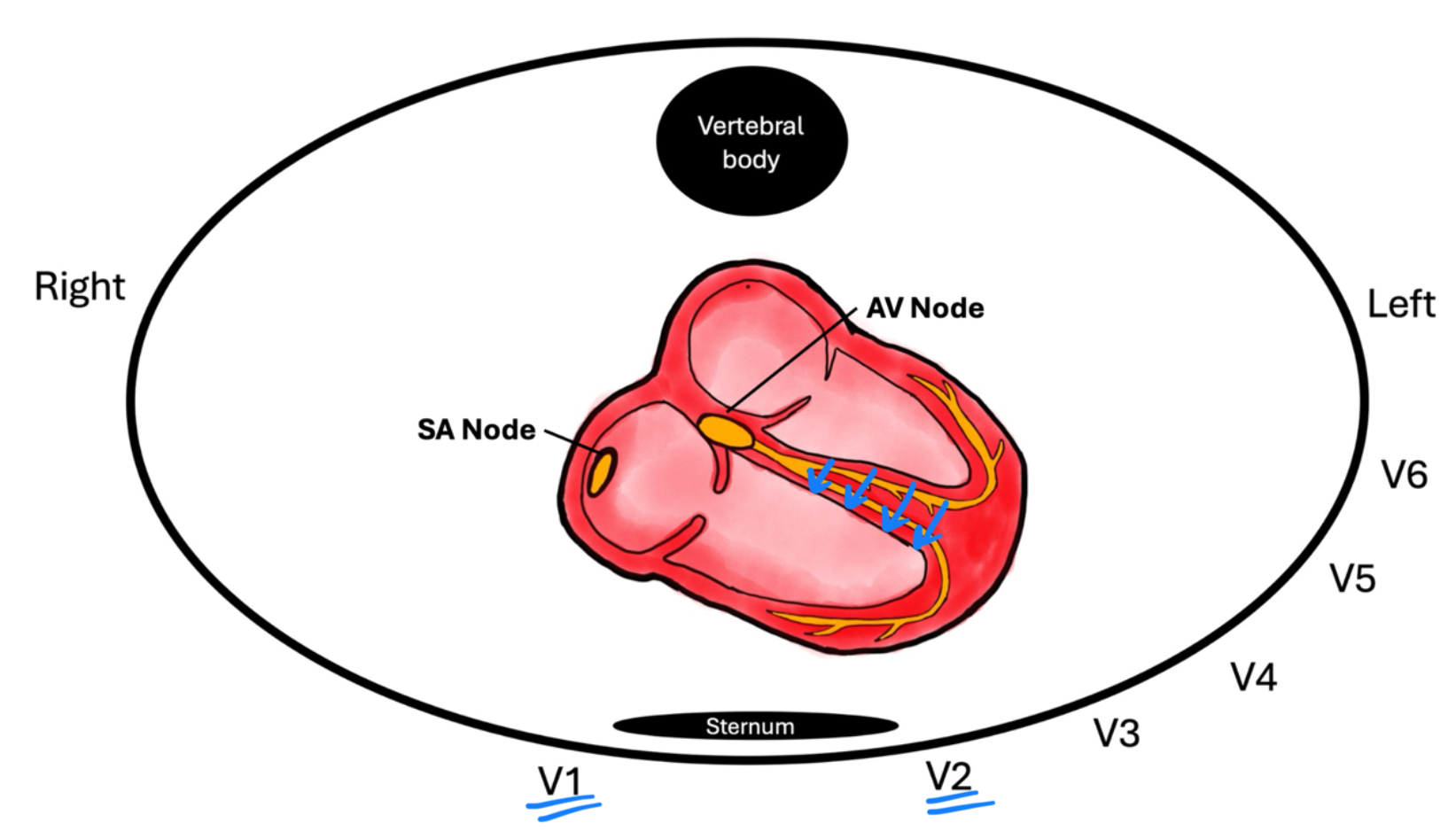
One of the subtle but critical features on the ECG is the septal R wave in the right precordial leads (V1 and V2). This small deflection often gets overlooked, but it reflects a very specific sequence of cardiac activation and provides important insight into normal conduction and anatomy. Understanding the septal R wave requires us to revisit the anatomy of the interventricular septum and the physiology of ventricular depolarization.
Related Topics:
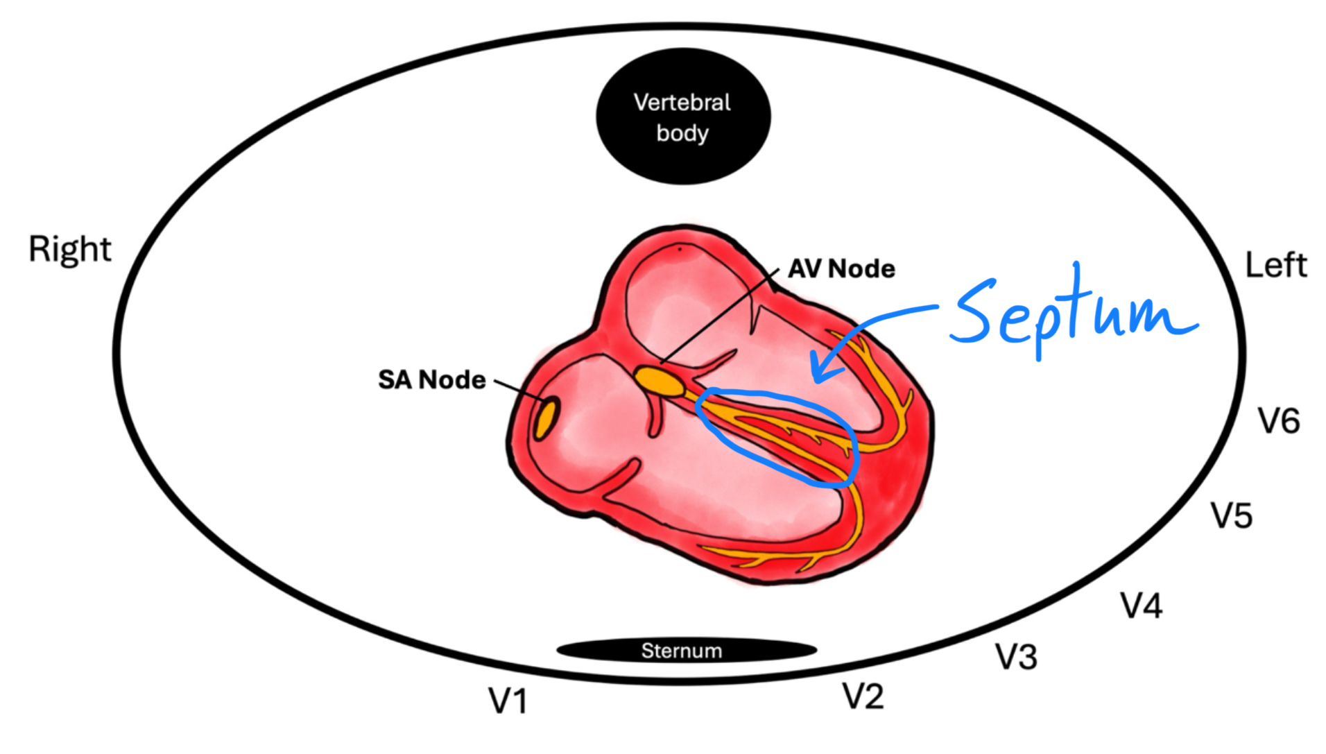
The Anatomy of the Interventricular Septum
The interventricular septum isn’t uniform—it has two distinct parts:
-
Muscular septum – the thick lower portion that makes up the majority of the septum.
-
Membranous septum – the thinner upper portion near the atrioventricular junction.
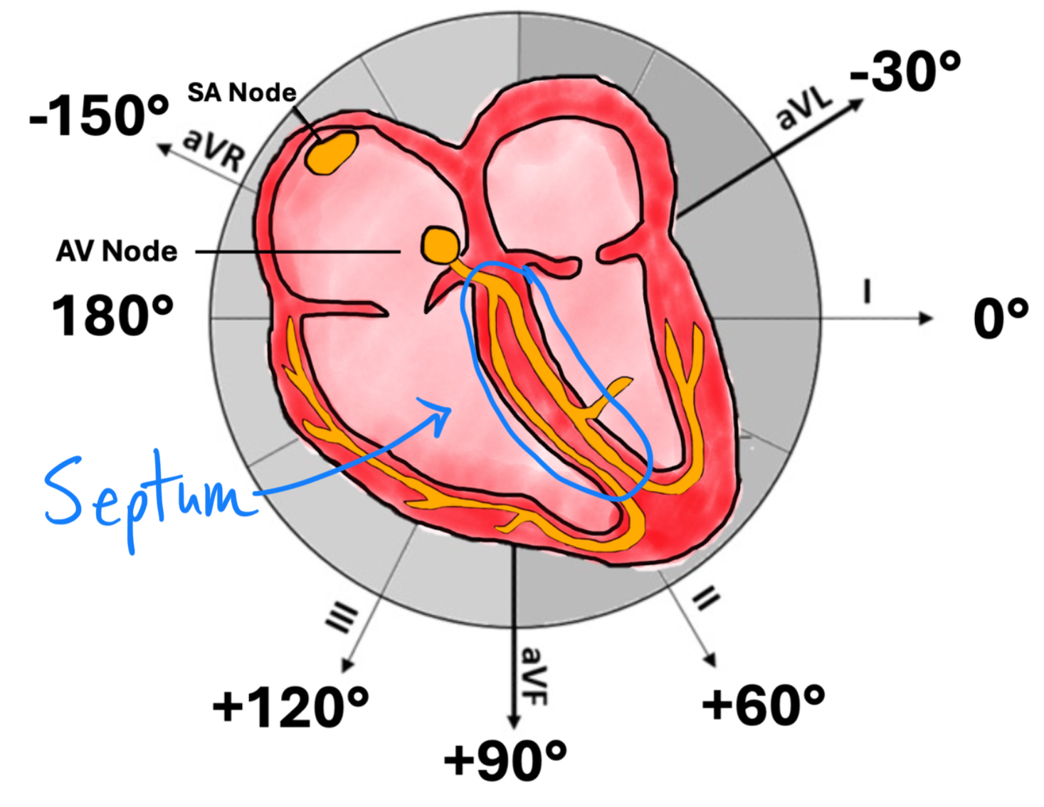
Embedded within the septum is the conduction system:
-
The bundle of His courses through the membranous septum before dividing.
-
The left bundle branch splits into anterior and posterior fascicles that spread across the left ventricle.
-
The right bundle branch runs down the right side of the septum.
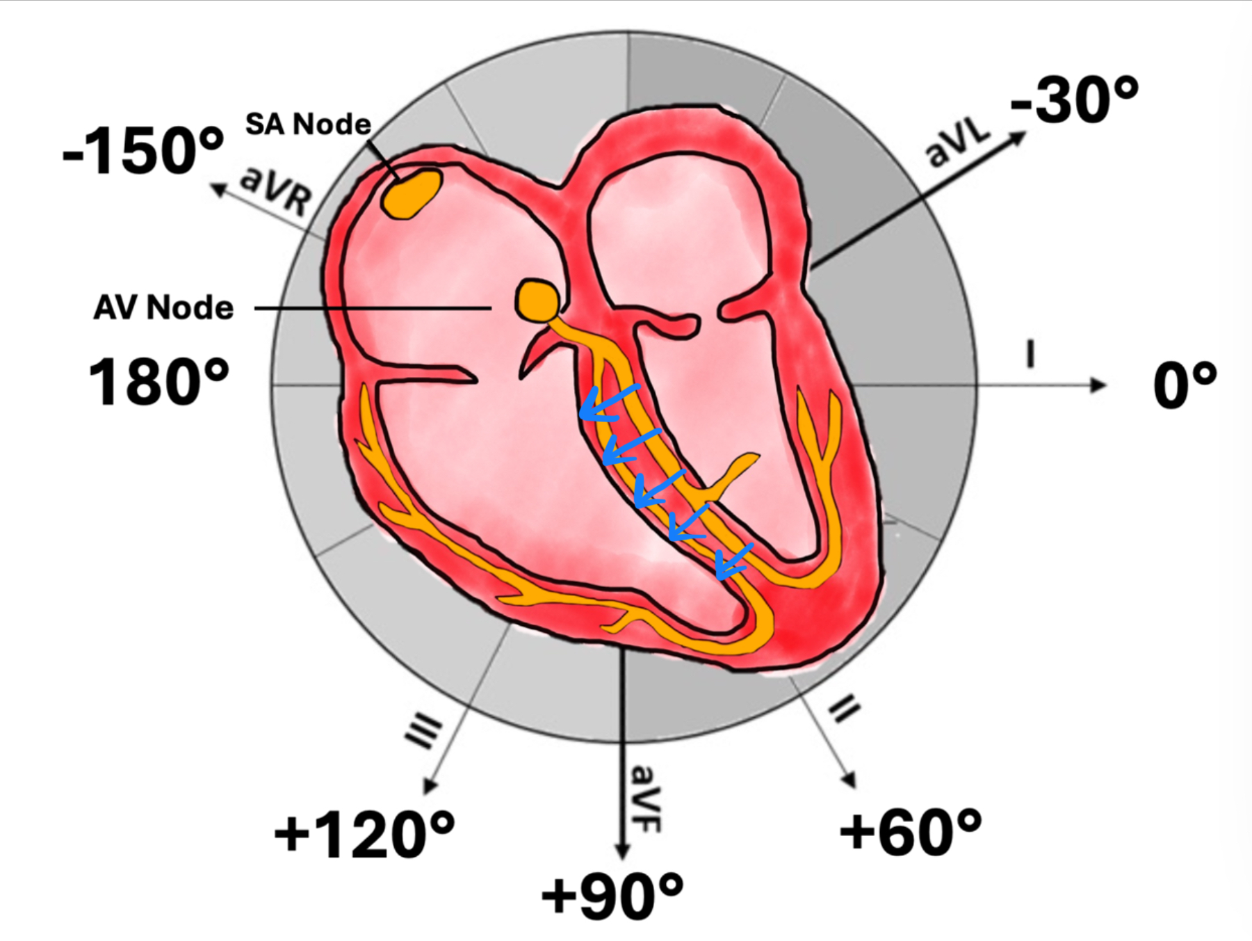
This anatomic arrangement explains why septal depolarization begins left to right, producing the small R waves in V1–V2.
Physiology of Septal Activation
Stepwise Depolarization:
-
Impulse from the His bundle enters the left bundle branch slightly before the right.
-
Septal depolarization begins left-to-right, activating the mid-septum toward the right ventricle.
-
This left-to-right vector is captured in the right precordial leads (V1–V2) as a small positive deflection: the septal R wave.
-
Almost immediately after, the large mass of the left ventricle depolarizes, producing a dominant S wave in V1–V2.

Key Concept:
The septal R wave is not about right ventricular activation—it’s about the earliest left-to-right depolarization of the interventricular septum.
ECG Appearance of the Septal R Wave
-
Leads: Seen in V1 and V2.
-
Size: Small, typically <3 mm in amplitude.
-
Duration: Narrow, as it reflects a brisk conduction across the septum.
-
Morphology: Appears as an rS complex (small r followed by deep S).
When the Septal R Wave is Abnormal
Loss or alteration of the septal R wave can be an important clinical clue:
-
Left Bundle Branch Block (LBBB)
-
The septum depolarizes right-to-left instead of left-to-right.
-
Result: Septal R wave disappears in V1–V2 (deep QS complex).
-
-
Anterior Myocardial Infarction
-
Infarction of the septum (often LAD territory) can eliminate the septal vector.
-
Result: Pathologic Q waves or absence of septal R waves in V1–V2.
-
-
Ventricular Pre-excitation (WPW)
-
Accessory pathway conduction may bypass the normal His-Purkinje sequence.
-
Septal activation becomes abnormal, altering or abolishing the expected septal R.
-
-
Right Ventricular Hypertrophy or Conduction Delay
-
May exaggerate or distort the rS pattern in V1–V2.
-
If you're interested, below is the link to my entire ECG Course
ECG With Reid Academy
Accredited by the AMA, AAPA, ANCC, ACPE & more.