
Fusion Beats in Monomorphic Ventricular Tachycardia
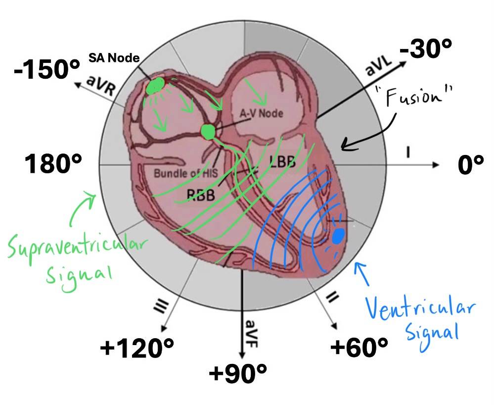
Jun 12, 2025Fusion beats are one of the hallmark findings that support the diagnosis of ventricular tachycardia (VT)—particularly in the case of monomorphic VT. They represent a unique moment in cardiac electrophysiology where two wavefronts—one ventricular and one supraventricular—collide, creating a hybrid QRS complex.
In this article, we’ll unpack the mechanism, morphological features, and clinical implications of fusion beats, and how they can help distinguish VT from supraventricular tachycardia (SVT) with aberrancy.
What Is a Fusion Beat?
A fusion beat is a hybrid QRS complex that results when two impulses reach the ventricles simultaneously from different sources:
-
One impulse arises from the ventricular focus (the ongoing VT)
-
The other is a sinus or atrial beat that briefly captures the conduction system and participates in ventricular depolarization
The result is a QRS complex that is intermediate in morphology between:
-
The pure VT QRS (wide, often bizarre)
-
A normal conducted beat (narrow QRS or with known baseline morphology)
Mechanism: Competing Activation
During monomorphic ventricular tachycardia, the ventricles are depolarized by a single ventricular ectopic focus, leading to:
-
Regular wide QRS complexes
-
AV dissociation (atria and ventricles depolarizing independently)
However, if the sinus node continues to fire independently—and one of those supraventricular impulses happens to reach the AV node and passes through the His-Purkinje system at the same time that a VT impulse is entering the ventricles, then:
-
Both impulses contribute to ventricular depolarization
-
The resulting QRS is a fusion of both wavefronts
-
The morphology is partially VT-like and partially normal, creating a “hybrid” complex
This only happens when the timing is just right: the supraventricular impulse must not be blocked at the AV node and must reach the ventricles during a window in which the VT impulse is also conducting.
ECG Morphology of a Fusion Beat
-
Occurs during a run of monomorphic VT
-
QRS complex is narrower than the VT beats, but still wider than normal
-
Morphology appears blended: part of the QRS resembles the VT beat; part resembles the patient’s normal QRS morphology
-
May occur as a single complex, or rarely in short series

Often, fusion beats are isolated and stand out in a regular sequence of identical VT QRS complexes.
Differentiating Fusion Beats from Other Wide Complexes
| Feature | Fusion Beat | Capture Beat | VT Beat |
|---|---|---|---|
| Origin | Dual: ventricular + supraventricular | Entirely supraventricular | Ventricular focus |
| QRS Morphology | Hybrid between VT and normal QRS | Normal or near-normal | Wide, bizarre, uniform |
| Occurs During | Monomorphic VT | Monomorphic VT | Monomorphic VT |
| Diagnostic Value | Strongly supports VT | Strongly supports VT | Supports VT, but not definitive alone |
Fusion beats differ from capture beats, which occur when a supraventricular impulse completely captures the ventricles, producing a normal QRS complex amidst VT—essentially a “breakthrough” beat.
Why Fusion Beats Matter Clinically
The presence of a fusion beat during a wide complex tachycardia is diagnostic of ventricular tachycardia. It confirms:
-
AV dissociation is occurring
-
The rhythm is not SVT with aberrancy, since the ventricles are not being exclusively driven by the atria
Fusion beats are one of the Brugada criteria for diagnosing VT and should raise confidence in the diagnosis when interpreting ambiguous wide complex rhythms.
Clinical Takeaway
Fusion beats represent partial depolarization by two competing wavefronts, one from the ventricles and one from the atria. When seen during a wide complex tachycardia, a fusion beat is powerful evidence in favor of ventricular tachycardia and helps distinguish it from SVT with aberrancy. Recognizing this pattern is crucial for accurate diagnosisand appropriate management, particularly in unstable patients.
Enjoy this style of lecture and looking to learn more? Click below to learn more about how you can join my complete ECG course.
ECG With Reid Academy
Accredited by the AMA, AAPA, ANCC, ACPE & more.
